
Radiology is that line of medical specialty which centers on contemplating, diagnosing and treating afflictions inside the human body using imaging. Since innovation has met quick evolvement, there are various methods that radiologists currently apply to get helpful images of within the human body. Radiology is a significant part of medical science which lets the radiologists or doctors to all the more likely evaluate what sickness is alarming the patient. It allows the medical experts to examine the patient’s body without cutting it open first. Through picturing the inner parts of the body onto a PC screen or an x-ray sheet, the radiologist will know for specific what sort of infection is affecting which organ definitively. A few specialty choices in diagnostic and restorative radiology incorporate chest radiology, stomach and pelvic radiology, body imaging, vascular radiology, mammography, pediatric radiology, outer muscle radiology and atomic medicine, just to give some examples. There is an entire bundle of imaging innovations that a confirmed radiologist can use for his potential benefit.
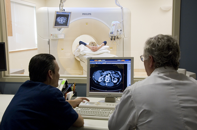
At the point when radiology was first presented quite some time ago, there were just x-rays and, surprisingly, that made a forward leap in the medical science at that point however for a long time, doctors just used plain radiology or evolved image on dull film which was empowered when a light emission rays are gone through a specific region of the patient. This sort of projection radiology is as yet being used even presently, as it is savvier and all the available. Also, this sort of imagery is more appropriate for the investigation of the skeleton, heart and bones. As the part of radiology expanded, so did the method for getting images from inside the body. After the typical x-rays which were caught onto a film, PC geography was presented this implied that now the images could be changed digitally and moved to a PC. So the radiologist could not directly see the images onto the PC screen.
A further developed type of diagnostic x-ray imagery is Fluoroscopy which consolidates the utilization of radio difference specialists, image intensifier tube and a fluorescent screen. It is likewise utilized for angiography. The image intensifier tube is connected to the PC framework through a shut circuit and click site https://prestigeer.com/services/imaging to read more information. The radio-contrast specialists can ingest x-rays and to dissipate them in this manner these specialists are either injected into the patient or given orally with the goal that they arrive at the inner parts of the body. When the x-rays are shot, an unmistakable ongoing image is gotten back to the TV in view of the radio-contrast specialists. Radiologists utilize this strategy when they need to concentrate on the blood stream in the veins and corridors or when they need to examine the genitourinary framework.